ARTHRITIS IN THOROUGHBRED RACEHORSES AND HOW TO PREVENT IT
—
When discussing the health and welfare of racehorses, always start by recalling the origins of their ancestors, thereby adapting their bodies to the environment in which they lived and developed. Horses are a grazed inhabitant of open savannas and are a typical example of a flight and fright animal. They have exceptionally well-developed locomotive system and mainly because of this, they have been serving mankind from around 3000 BC. Every change introduced to their natural existence may result in body diseases and injuries of the locomotor system.
 |
The racing world is one of many that takes advantage of the mobility of horses and therefore often exceeds their endurance. Musculoskeletal disorders, especially arthritis of the carpal and fetlock have become a very common problem and rank as first among the causes of waste in the racing industry (Rossdale et al., 1985, Bailey et al., 1999, Wilson et al., 1996, Church et al., 2009). In addition, veterinary medicine, despite its efforts, does not currently offer a cure for arthritis.
Arthritis is a joint inflammation. Like any joint, which is simply a connection between two bones, inflammation can incite a cascade of events that ultimately leads to long term uncontrolled inflammation, called osteoarthritis, the effects of which are irreversible and depending on the degree of development may lead to lameness and poor performance of horses. All types of arthritis are defined as painful, leading to stiffness and permanent degeneration of the cartilage in the horse's affected joint. In adult horses, it may take several weeks or months for the first signs of secondary osteoarthritis to appear. This situation leads to progressive and permanent loss of articular cartilage and changes in the bone itself, joint capsule and synovial membrane (Hunzike, 2001).
Therefore, it is very important to know all the factors that can contribute to the development of arthritis in order to minimize the risk of the disease, and give the horse the best possible welfare and finally maximise racing longevity and the best life after racing career. There are relatively few studies investigating the risk of arthritis and focusing on prevention of the Thoroughbred (Corbin, et al., 2012). For this reason we face the challenge of making our science follow this path as well. For this reason we face the challenge of making our science following this path as well. To understand the prevention or minimisation of development of osteoarthritis among racehorses, first understand the factors that may contribute to its development. Many things can stress a joint. However, the best recipe for racehorses arthritis is repetitive, rigorous training connected with long-term bad nutrition and poor hoof conformation.
Let's take a closer look at these three subjects.
TRAINING
To understand the effect of training on development of arthritis, the joints should be seen as organs where the constituting tissues like articular cartilage, subchondral bone, synovial membrane, synovial fluid, intra-articular ligaments and menisci act together. Proper joint homeostasis is the key element, and any pathological changes within individual elements affect the work and health of the entire joint. Race horses are highly prone to arthritis at any age. This is because they are already subjected to rigorous and repetitive training before they reach physiological maturity. The thoroughbreds are broken in as a yearling, and trained and raced as a two years old. It is well known that horses which are broken too early can wind up having joint problems and soundness issues as they age. Often, the only determinant for starting training is the end of the carpal joint union. The unfinished growth of the carpal joint almost always guarantees injuries, and hence exclusion from the racing career (Kawcak, et al., 2000). However, it was already shown in the 1970s that objective measures, such as physeal (growth plate) closure of the radial bones, were generally ineffective in assessing musculoskeletal maturity and suitability for racing (Gabel, et al, 1977).
Monotonous overload training expose the joints to stress. It has been reported repetitive or sudden mechanical impact during exercise is a significant risk factor for joint abnormality, and the equine carpal osteochondral fragment combined with exercise is a predictable and well-accepted model for arthritis (Davidson, 2016). On the other hand, lack of exercise causes a lack of lubrication in the joints and its poor development, which may contribute to the development of inflammation at an early age of the horse. In young, growing individuals the plasticity of cartilage is considerably greater and this makes that discrimination should be made between the effects of exercise in full-grown adults and juvenile individuals. The cartilage of a young, immature horse is soft, still forming and cannot withstand intense or repetitive work. Healthy joints are protected by cartilage, which acts as a cushion between the bones, and a thick, sticky synovial fluid that acts as a lubricant. Stress in the joint cannot be prevented, but the goal is to normalise the joint inflammation as quickly as possible, before permanent damage occurs. As the horse moves, flexion and pressure can cause minor damage to the joint structures that trigger mild inflammatory responses to repair. Usually, the body's defences control inflammation and the joint remains healthy. Repeated and rigorous training overwhelms the body's ability to contain it, whether from a single acute injury or after many years of use. Inflammatory enzymes break down the lubricating synovial fluid, which becomes thinner. Proteoglycans are lost and collagen fibres lose their integrity, reducing the cartilages ability to retain moisturising fluid. This damage stimulates even more inflammation that fills the joint capsule with fluid, leading to pressure, pain and stiffness. The build-up of inflammatory enzymes further breaks down the synovial fluid, leading to more damage of the cartilage. Progressive subchondral bone sclerosis leads to thinning of articular cartilage. On the other hand, primary loss of articular cartilage leads to subchondral bone sclerosis. The progressive nature of the changes in the joint at the osteochondral junction leads to pain. Some horses may not show any signs of lameness, but there may be swelling, filling, heat or pain around the affected joint. These are all common symptoms of arthritis.
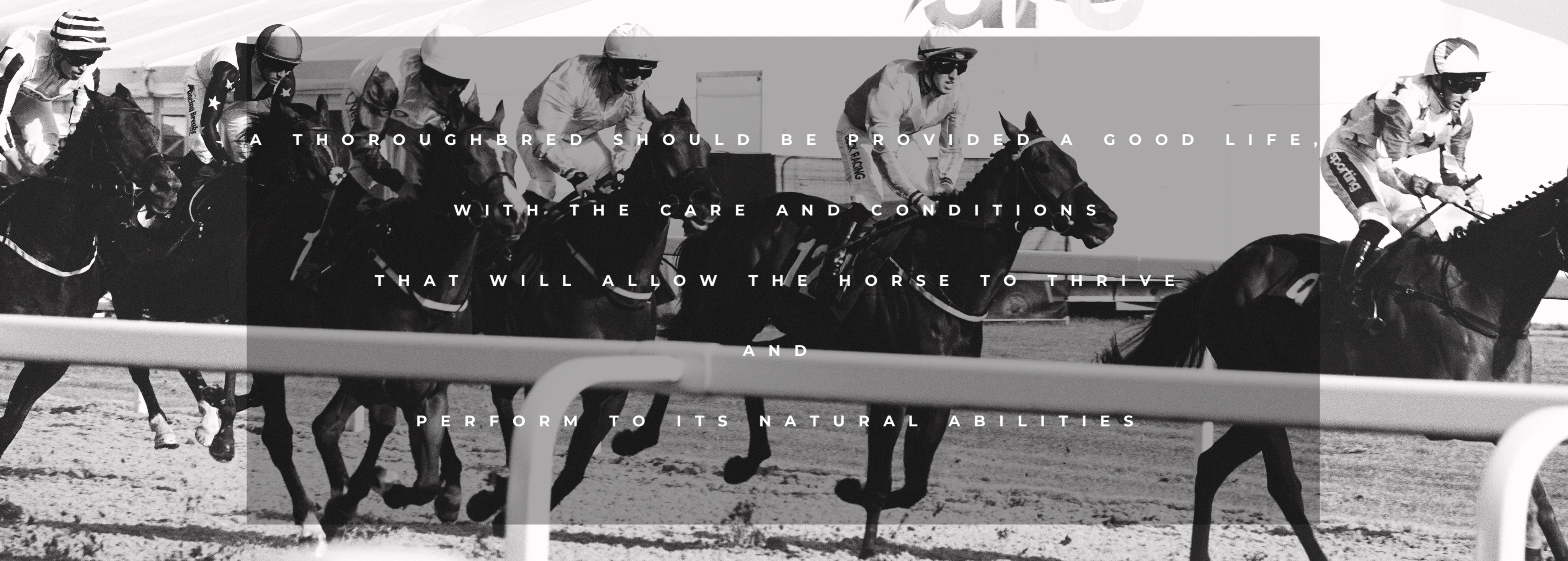
The long-term effect of training on joints is nothing but changes in mechanical loading of joints, which elicit responses from the tissues and hence affect bone homeostasis repeated in period of time. With an optimal training program, disturbed homeostasis has a chance to return to normal during the rest period, thanks to which the individual elements adapt to exercise. If the limit is drastically exceeded (direct trauma) or repeatedly overloaded (fatigue stress), instead of adoption, permanent pathological changes occur. Also, asymmetry of the rider as an additional imbalance weight may have negative influence on joints health through incorrect load distribution (MacKechnie-Guire, et al., 2020).
JOINT NUTRITION
The fundamental of healthy bones and joints is their proper nutrition. The nutrients such as minerals and trace elements play important role in bone development during growth and in the maintenance of bone mass thereafter. Poor nutrition contributes to a faster breakdown of joint homeostasis increased susceptibility to arthritis during training and racing, and decreased ability of the body to regenerate damaged cells. Long-term deficiencies, excesses, and imbalances of nutrients may result in breakdown joint homeostasis, weakening of joint structures, an increase in the incidence and severity of arthritis. In practice, in the diet of racing horses, we mainly deal with a deficiency and imbalance rather than an excess of minerals in the diet. Most commonly fed cereal grains and forages contain insufficient quantities of several minerals. Additionally, the lack of an appropriate worming program increases the probability of worms in the digestive system, which remove the nutritional value of wormed horses. Let's take a look at the most important building and homeostatic element in the joints.
CALCIUM & PHOSPHORUS
Calcium (Ca) and phosphorus (P) are generally considered a key element for maintaining bone mineral homeostasis. Calcium is the basic building block of bones. Long-term calcium deficiency in the body leads to chronic soreness of muscles and joints, degeneration and diseases of bones and joints. Bones become brittle, weak and prone to breakage. The major function of phosphorus is in the formation, with calcium, of the bone component hydroxyapatite. 80% of the phosphorous in the body is present as calcium salts in the skeleton and, therefore, is essential for healthy bone. Ca and P may be required to support the remodeling of bone associated with exercise in young racehorses. Ca and P also may be indicated during recovery after intense exercise and during period of inactivity (Nielsen, 2013). High levels of phosphorus inhibit calcium absorption and lead to a deficiency, even when the amount of calcium present is usually adequate. The diet, which contains too much grain and too little forage, is high in phosphorus and low in calcium (Gruenberg, 2019). However, too much calcium can affect the phosphorus level, especially if the phosphorus level is negligible.
 |
SODIUM
Sodium (Na) is responsible for regulating body water content and electrolyte balance. Sodium is also required for the absorption of certain nutrients and water from the gut. It is known that high sodium intake in the diet reduces calcium reabsorption in the kidneys, which in turn leads to greater excretion of calcium in the urine. This effect has been demonstrated in studies on humans of all ages, as well as in experimental animals (Saric and Piasek, 2005).
POTASSIUM
Potassium (K) is essential for water and electrolyte balance and the normal functioning of cells.
MAGNESIUM
Magnesium (Mg) has important interrelationships with calcium, potassium and sodium. It is needed for parathyroid hormone secretion, which in involved in bone metabolism.
FLUORIDE
The main function of fluoride (F) in the body is in the mineralisation of bone.
COPPER
Copper (Cu) is the third most abundant dietary trace metal after iron and zinc. The body needs copper to utilise iron efficiently and it is thought to be important for foal growth and for strong bones. Copper deficiency may be due to an excess of zinc in the body.
MANGANESE
Manganese (Mn) is required for bone formation.
CHLORIDE
Chloride (Cl) ions play a major role in osteoclast biology and bone homeostasis.
VITAMIN D
Vitamin D is another very important building and homeostatic element in the joints. Vitamin D is obtained either through the diet or by exposure to sunlight. If the vitamin is decreased, calcium and phosphorus absorption are reduced. Bone deformities can result, as well as other nutritional and metabolic complications. Photo 2. 5-year-old gelding with osteoarthritis with visible rickets on the nasal bone, which indicates a long-term lack of bone building elements (Vit.D, Calcium). Rickets may be initially caused by a calcium deficiency alone (hypocalcemia) due to low dietary calcium intake. Low calcium levels increase vitamin D utilization and may decrease vitamin D levels, resulting in a combination of calcium deficiency and vitamin D deficiency or rickets failure. [1] Bones are a reservoir of calcium. In case of inadequate blood level due to the need to maintain adequate blood levels, the effect of an inadequate calcium supply is usually reflected in bone density as bone acts as a reservoir when needed. Insufficient amount of calcium in the bones may be due to insufficient supply of vitamin D, which is essential for calcium absorption. In foals, vitamin D deficiency leads to rickets, which affects the growth plate in growing horses. However, in adults horses, to osteomalacia, in which the bones become weak due to a lack of calcium.
SWEATING
Taking care of the quality of the joints, one must not forget about supplementation balance, taking into account the loss of micronutrients important for the joint through sweat excreted during training. Sweat includes 98-99 percent water, and salt, fats, urea, ammonia, lactic acid, minerals. Sodium, potassium, calcium and magnesium are elements whose proper balance is very important for maintaining the correct water and electrolyte balance of the body. Horses mainly lose them with sweat. Therefore, it is important to replenish these losses by drinking water and consuming foods rich in the appropriate elements. In comparison to humans, peak sweating rates in humans are ~3 L / hour compared to ~20 L / hour in horses (Lindinger, 2008). Effective prevention of extracellular dehydration requires Na, K, CL, Ca and Mg in electrolyte supplements. In theory, imbalance these elements promote the demineralization of bone as well as increased renal Ca loss (Baker, et al., 1993, 1998). These elements are also important for cellular function. Sweat contains a small amount of iron and other trace element (e.g. zinc), there may be a slightly increased requirement with exercise.
HOOF CONFORMATION AND BALANCE
When discussing the impact of training on developing of arthritis, one cannot fail to mention the appropriate balance of the horses' legs, on which the forces during landing must be optimally distributed evenly over the entire surface and length of the leg. Balance is the term used to describe the relationship between the horse's limb, foot and shoe. When the balance during loading is disturbed, articulating surfaces, loaded inappropriately, and increased risk of osteolytic changes is made. Long toe, low heel and more horizontal landing forces are typically for racehorses. Poor conformation of the hoof causes poor balance and bad distribution of forces on the legs, which lead to move more stress on the individual components. However, we have no influence on genetics, we are able to try to support horses to cope with demands of workload during training and racing. It is extremely important to find a farrier that can not only correct and keep the hoof in balance, but can also balance the hoof to the conformation of the limb. X-ray imaging are very helpful in the diagnosis, unfortunately it is very expensive and considering the large number of horses in training, not profitable for the owners. The visual assessment of the angles may turn out to be inadequate to the actual angles of the skeletal system. This means that by trimming the hoof evenly, you can curve the joint, causing the joint to be unevenly stressed, which can degenerate the joint components. Even small imbalances may have considerable biomechanical effects (Dippel, et al., 2016). The vet-farrier relationship is very important to provide the best management and prevention to avoid injuries of the joints.
MANAGEMENT OF NON-OPERATIVE ARTHRITIS
—
As it was mentioned in the beginning, this disease can not be cured, but by appropriate management, we can slow down progression of this condition. Catching the disease early is the key to effectively slow down the ongoing damage and managing accompanying pain. Arthritis management offers using of medication, supplements and physiotherapy. It is varied based on the severity of disease, the severity of compensatory pain and the individual horse’s pain tolerance.
MEDICATION
Medications are included NonSteroidal Anti-Inflammatory Drugs (NSAIDs), systemic hyaluronic acid (HA), polysulfated glycoaminoglycan (PSGAG), and corticosteroids. Consequently, combination of these medication is often used. It should be mentioned that the most frequently administered NSAIDs agents are associated with side effects with long-term use. The most frequently reported adverse effects are gastrointestinal side effect. Gastric mucosa degeneration progressed with the duration of NSAID treatment. (Nicpon, et al., 2000). Many race horses struggle with peptic ulcer disease, and for this reason, orally administered medication becomes limited or difficult. A good solution may be topically applied liposomal diclofenac sodium cream (LDS) (Surpass), which has beneficial disease modifying effects (Hardman, et al., 1996, Frisbie, et al.2009, Javeed, 2011). In addition, interleukin-1 receptor antagonist protein (IRAP) has been shown to decrease pain and reduce cartilage degradation (Frisbie, et al.2002). The polyacylamide hydrogel, which is use in urology and plastic surgery in humans, shows promise as a treatment for arthritis in horses (Tnibar, et al, 2015).
SUPPLEMENTATION
Support therapy like supplements can be use in long term administration. Glucosamine, chondroitin sulfates, hyaluronan, pentosan polysulfate can be found naturally in joints. Glucosamine is believed to play a role in the formation and repair of cartilage, chondroitin sulfate helps give cartilage its elasticity, and hyaluronan helps lubricate joints and form the matrix of articular cartilage. Some newer supplements contain soybean unsaponifiables (ASU) extracts and avocado, which studies suggest may reduce inflammation and protect cartilage by increasing glycosaminoglycan (GAG) synthesis (Kawcak, et al, 2007). Also, many publications have shown beneficial effect of various herbs and natural anti-inflammatory like turmeric, fish oil, arnica, quercetin, green tea, curcumin, boswellia serrata, frankincense essential oil on intervention for cartilage inflammation and pain relief. Example, Singht et al (1997a) administrated orally herbal preparation containing Allium sativum (garlic), Cyperus rotundus (nut grass), and Zingiber officinale (Ginger) in donkeys with aseptic arthritis. In treated animals, changes in the joint capsule, synovial membrane and articular cartilage were far less than in untreated animals. The changes were restricted to tangential layer only. When discussing supplementation, the importance of regular deworming should be taken into account. Wormed horses will not absorb nutrients as much as possible and will also be poisoned with worm droppings, which adversely affect the joints.
NON INVASIVE THERAPEUTIC TREATMENT
Role of the non invasive therapeutic treatments is to optimize natural healing process. Physiotherapy offers manual therapy and physio devices, which work witch biomechanical and biochemical level by putting energy into the tissue to change it behaviour, and achieve physiological effect.
ASSESSMENT
Regular assessment can early detect of mild changes and pathology in the joints, which can help to preserve the career or indeed life of many race horses. Subchondral bone is, unlike cartilage, richly vascularized and has very well developed nerve supply that plays a major role in pain perception in joint disease. Because of that palpating these areas can be helpful for daily assessment. Identification of the ‘at-risk horse’ rather the identification of risk factors, can allow to plan appropriate prevention, which will allow to extend the racing career and the horse's well-being. Also, daily checking of the legs for increased heat by the trainer is extremely important from the preventive point of view. This allows for quick detection of clinical signs of pathological changes and early intervention before the horse begins to compensate for the pain and then manifest it lameness.
MANUAL THERAPY/ JOINT MANIPULATION AND MOBILIZATION
Joint manipulation and mobilization can be provided as an assessment and therapeutic approaches in veterinary physiotherapy. It allows the examination and assessment of the quality and range of joint motion to detect abnormalities. Also, it reduces the risk of joint inflammation or delay its onset by supporting and maintaining the health of horse’s joints. Therapeutically, these techniques can help reduce pain and decrease swollen joints, treat secondary compensation, and maintaining homeostasis of the joints for the future. In case of arthritis the primary aim of the therapeutic management is to return the joint to non painful function and limit the formation and progression of osteoarthritis. Also, it is important to help rapid resolution of synovitis and capsulitis to avoid capsular edema and capsular fibrosis that may lead to reduced ROM of the joint (Pamer, and Bertone, 1996, Pool, 1999). Race horses are often stalled during the training period without being allowed to move freely in the pasture. However, training sessions take place mainly under the rider, which also restricts the freedom of movement. These limitations can disrupt the natural manipulation of the joints. This leads to cascading changes in the musculoskeletal system that can affect joint quality. However, joints, which are subjected to a controlled opposing force can be kept in a much better condition than those that are used only in a single direction. Haussler explains in many scientific works that spinal manipulation is effective to reducing pain, improving flexibility, reducing muscle tone, and improving symmetry of spinal kinematics in horses (Haussler, 2016).
THERMOTHERAPY
Thermotherapy is a very accessible and easy way to prevent and treat arthritis. Cold and heat are used separately or alternately depending on the case and intended purpose. Cryotherapy is provided to control inflammation by providing anti-inflammatory analgesic effect, reduce cellular metabolic demands, decrease pain, induce muscle relaxation. Therapeutically, it should be used in acute injuries until inflammation is resolved (48-72h), and in chronic cases, after exercises. Heat therapy can provide increased soft tissue extensibility, reduced inflammation and adhesion formation, pain control to help facilitate the restoration of normal joint motion. Affected horses avoid joint movement due to pain. Used before exercise, it warms up the muscles and stiff joints, thus preparing them better for exercise while minimizing any discomfort or pain. Also, when used overnight, it allows body to optimize the renewal process. Routine management procedures such as icing and thermal bandaging should be provided and promoted in every racing yard, both while training season and also resting and rehabilitation interval.
THERAPEUTIC ULTRASOUND
Equidae with aseptic arthritis in the carpal joint treated with pulsed ultrasound device returned early to normal stance and weight bearing than control animals (Singh, et al., 1996). Synovial total leukocyte count (TLC) decreased as the treatment started. However, TLC in untreated group increased progressively. Ultrasound also decreased synovial total proteins (STP) to near normal levels where remained so at all subsequent intervals in the control group. Synovial and plasma lactate dehydrogenase activity was found significantly increased in control group and non significant change was observed in treated group. The gross changes in the joint capsule, synovial membrane and articular cartilage were quite mild in ultrasound treated animals compared with controls (Singh, et al., 1997c). In another study, Singht shown that the joint capsule in the treated animals by ultrasound was almost normal and there was no calcium deposition after 7 days of treatment (Singht, et al., 1997b). Also, While, degeneration of the articular cartilage was observed in untreated animals. It is concluded that ultrasound therapy is an effective treatment for arthritis in equidae. Ultrasound therapy resulted in satisfactory healing of joint tissue and articular cartilage and therefore presented the degenerative joint disease observed in untreated animals. More information about therapeutic ultrasound you can find here.
THERAPEUTIC PEMF
Pulsed electromagnetic field therapy has been proven to be safe, tolerable and an effective treatment for arthritis and to slow down degenerative changes. Researches have shown that long term imbalance between the synthesis and decomposition of chondrocytes, cell matrices and subchondral bone, leads to the degeneration of articular cartilage. PEMF stimulate proliferation of chondrocytes and exert a protective effect on the catabolic environment It decreases pain, inflammation and get a more normal cellular environment. More information about therapeutic PEMF you can find here.
THERAPEUTIC EXTRACORPOREAL SHOCK WAVE
Extracorporeal shock wave therapy, which involves directing a beam of energy waves at a target site, has shown promise as a treatment for osteoarthritis in horses. In one study, treated horses showed significant improvement in clinical lameness as well as in the concentrations of certain biochemical markers of the disease. However, some research has been shown no disease modifying effect (Frisbie, Kawcak, McIlwraith, 2009, 2011).
THERAPEUTIC EXERCISE ON THE GROUND
Therapeutic exercise is a fundamental part of rehabilitation program for arthritis horses. Exercise helps keep joints healthy by stimulating the production of synovial fluid and by strengthening the muscles that help stabilize the joints. Program aims to return the joints and affected soft tissues to normal physical capacity, and in long term goal strengthening and maintaining of joint mobilizers. It has been reported the therapeutic effects of exercise on cartilage lesions include thicker repair tissue, increased glycosaminoglycan content, and less bone remodelling with exercise (French, et al, 1989, Foland, et al, 1994). To understand the basics of a suitable exercise program, knowing normal tissue response after damage is essential (more about healing process you can find here). However, bone has the amazing ability to heal itseal, cartilage has a limited ability to heal (Hunzike, 2001). Poor conduct in the rehabilitation process, such as allowing the horse to return too quickly or suddenly to free, uncontrolled movement increases the likelihood of returning to an injury or weakening the pattern of the renewal process, which may result in weaker tissue reconstruction. Horses with arthritis on closer inspection reveal multiple related or unrelated problems. The goals are to keep them as active as possible, in order to stimulate circulation, while minimizing the risks of overuse and the inflammation it brings. The exercises must be selected individually, because many factors affect the planning of the program, such as the type of arthritis, its advancement, age and condition of the animal, as well as environmental possibilities. Adjustments should be made to properly prepare a musculoskeletal for work in cold weather. Tailor warm-up to the individual horse’s needs and preferences. On the other hand, the lack of free movement access during the growing up period from birth to the start of training leads to poor musculoskeletal development, which can contribute to faster wear of joints during the rigorous training period of racing horses. So, it is very important to optimize turnout of foals and weanlings as much as possible. An important element in reducing the risk of arthritis is the proper preparation of the body to the load. The tendons and ligaments can tear, and the joint can easily be dislocated if the body is not warmed up properly before strenuous exercise.
THERAPEUTIC EXERCISE IN THE WATER
Water is a very beneficial element in the prevention and treatment of arthritis in racing horses. Underwater treadmill and swimming have been reported to decrease pain and inflammation, improve joint range of motion, and strengthen the surrounding tissues (Kamioka, 2010). It provides an effective practice for promoting normal motor patterns, increasing muscle activation, and reducing the incidence of secondary musculoskeletal injuries caused by primary joint pathology (Prins, 1999). Exercising patients in the water may be beneficial before starting flat work to increase the strength of the tissues without overload joints.
DIGRESSION
 |
Along with the using of Thoroughbreds in racing industry, we are obligated to ensuring their best welfare. However, often the most important is economic aspect of the business than well-being of animal. Also, overbreeding in this specific industry contributes to the superficial treatment of the welfare of horses and treating them primarily in the economic aspect. Of course economic restraints also apply when considering the management of equine arthritis. This can be particularly problematic when clients realise that vets are unlikely to be able to cure the condition and that therapy may be required for a number of years. In the present economic climate the diagnostic work up and conservative management are very expensive and may cost in excess of the monetary value of the horse. In this case, the fact that limiting comments to lesions and conformational faults only, and their likely effects on future athletic soundness appears the most appropriate advice for the owners. Therefore, the breeding program should be more sensible, including the selective selection of mares for breeding. If you look at the problem of arthritis in horses from a prophylactic point of view, prevention can also turn out to be economically beneficial. Preventive management programs may allow early detection of joints disease, therefore prevent or minimize the high cost of vet treatment, prolong racing longevity, and most importantly, provide the best care during training and also life after racing. This is why prophylactic management to prevent and minimize risk of arthritis and related with arthritis diseases should be basic in a good stable management. Continuity and regular assessment of the individual horses by physiotherapist may allow early detection of joints disease. In prolonging racing longevity alternative, combined training programme can be the best decision. It can include lower-intensity training like swimming, longer intervals between high-speed work combined with intermittent anti-inflammatory physical therapy and continual use of disease-modifying arthritis agents and supplements. Also it is well known that proper nutrition and maintenance of joint health takes place only when the joint is in motion, balancing this movement is the key to preventive treatment against the development of arthritis.
- Because Prevention is the Best Policy -
References:
Bailey CJ, Reid SW, Hodgson DR, et al. Impact of injuries and disease on a cohort two and three years old Thoroughbreds in training. Vet Rec 1999; 23: 487-93.
Baker L.A., Topliff D.A., Freeman D.W., et al. Effect of dietary cation-anion balance in mineral balance in anaerobically exercised and sedentary horses. J Equine Vet Sci 1993;13:557-61.
Baker L.A., Topliff D.A., Stoecker B. The comparison of two forms of sodium and potassium and chloride versus sulfur in the dietary cation-anion difference equation: Effects on acid-base status and mineral balance in sedentary horses. J Equine Vet Sci 1998;18:389-95.
Brown M.P., Trumble T.N., Plaas A.H., et al. Exercise and injuryincrease chondroitin sulfate chain length and decrease hyaluronan chain length in synovial fluid. Osteoarthritis Cart, 2007; 15:1318-25.
Corbin L.J., Blott S.C., Swinburne J.E., et al. A genome-wide association study of osteochondritis disseacans in the Thoroughbred. Mamm Genome 2012; 23:294-303.
Church E.E., Walker A.M., Wilson A,M., et al. Evaluation of discriminant analysis based on dorsoventral symmetry indices to quantify hindlimb lameness during over ground locomotion in the horse. Equine veterinary journal, 2009; 41,3,304-8.
Davidson E.J. Controlled exercise in equine rehabilitation. Vet Clin Equine 2016;32:159-165.
Dippel M., Ruczizka U., Valentin S., et al. Influence of increased intraarticular pressure of the isolated equine distal interphalangeal joint. J Equine Vet Sci 2016;38:54-63.
Foland J.W., McIlwraith C.W., Trotter G.W., et al. Effect of betamethasone and exercise on equine carpal joints with osteochondral fragments. Vet Surg 1994;23:369-76.
French D.A., Barber S.M., Leach D., et al. The effect of exercise on the healing of articular cartilage defects in the equine carpus. Vet Surg 1989;18:312-21.
Frisbie D.D., Ghivizzani S.C., Robbins P.D., et al. Treatment of experimental equine osteoarthritis by in vivo delivery of the equine interleukin-1 receptor antagonist gene. Gene Ther 2002;9:12-20.
Frisbie D.D., Kawcak C.E., McIlwraith C.W. Evaluationof the effect of extracorporeal shock wave treatment on experimentally induced osteoarthritis in middle carpal joints of horses. Am J Vet Res 2009;70:449-54.
Frisbie D.D., McIlwraith C.W., Kawcak C.E., et al. Evaluationof topically administered diclofenac liposomal cream for treatmentof horses with experimentally induced osteoarthritis. Am J Vet Res 2009;70(2):210-15.
Gabel AA, Spencer CP, Pipers FS. A study of correlation of closure of the distal radial physis with performance injury in the Standard bred, J Am Vet Med Assoc 1977; 170(2): 188-94.
Grace N.D., Pearce S.G., Firth E.C., Fennessy P.F. Concentrations of macro- and micro-elements in the milk of pasture-fed thoroughbred mares. Aust Vet J. 1999;77(3):177-180.
Gruenberg W. Disorders Associated with Calcium, Phosphorus, and Vitamin D in Horses. 2019 https://www.msdvetmanual.com/
Haussler K.K, Joint Mobilization and Manipulation for the Equine Athlete. Vet Clin North Am Equine Pract. 2016; 32(1):87-101.
Hardman J.G., Limbird L.E., P.B. Molinoff, R.W. Rudden. (1996) Goodman and Gillman’s The Pharmacological Basic of therapeutics. Maxwell MacMillan Int.Ed., pp.618, 637.
Hunzike E.B. Articular cartilage repair: basic science and clinical progress. A review of the current status and prospects. Osteoarthritis cartilage 2001;10:432-63.
Javeed A. (2011) Diclofenac sodium and equine arthritis.
Kamioka H., Tsutanji K., Okuizumi H., et al. Effectiveness of aquatic exercise and balneotherapy: a summary of systematic reviews based on randomized controlled trials of water immersion therapies. J Epidemiol 2010;20:2-12.
Kawcak C.E., McIlwraith C.W., Norrdin R.W., et al. Clinical effects of exercise on subchondral bone and metacarpophalangeal joints in horses. Am J Vet Res 2000; 61:1252-8.
Kawcak C.E., Frisbie D.D., McIlwraith C.W., et al. Evaluation of avocado and soybean unsaponifiable extracts for treatment of horses with experimentally induced osteoarthritis. Am J Vet Res 2007; 68: 598-604.
Kawcak C.E., Frisbie D.D., McIlwraith C.W., et al. Effects of extracorporeal shock wave therapy and polysulfated glycoaminoglycan treatment on subchondral bone, serum biomarkers, and synovial fluid biomarkers in horses with induced osteoarthritis. Am J Vet Res 2011;72:772-9.
Lindinger M. Sweating, dehydration and electrolyte supplementation: Challenges for the performance horse. Proc. of the 4th European Equine Nutrition & Health Congress April 2008; 46-56.
MacKechnie-Guire R., MacKechnie-Guire E., Fairfax V., et al. The effect that induced rider assymetry has on equine locomotion and the range of motion of the thorocolumbar spine when ridden in rising trot. J. Equine Vet Sci. 2020;38:
Nielsen B.D. Practical considerations for feeding racehorses. In: Geor R.J., Harris P.A., Coenen M.C., editors. Equine Applied and Clinical Nutrition. London: Saunders Elsevier; 2013. p. 261-71.
Nicpon, J. K., Kubiak K., Dzimira S., Sapikowski G. (2000). zeffect of selected non-steroidal anti-inflammatory drugs (NSAIDs) on canine gastric mucosa. Medycyna-Weterynaryjna, 56(9):582-5.
Palmer JL, Bertone AL. Joint biomechanics in the pathogenesis of traumatic arthritis. InMcIlwraith CW, Trotter GW, editors. Joint disease in the horse. Philadelphia: WB Sauders; 1996. p. 104-19.
Pegan J.D.The role of nutrition in developmental orthopaedic disease: nutrition management. In: Ross M.W., Dyson S.J. editors. Diagnosis and management of lameness in the horse. Saunders Elsevier; 2011; 2nd, p. 625-631.
Perkins N. Wastage in NZ Thoroughbred racing industry: an epidemiological investigation. In Proceeding Annual Seminar Equine Branch New Zealand Vet Association. 1999. p 103-12.
Pool RR, Pathologic manifestations of joint disease im the athletic horse. In: McIlwraith CW, Trotter GW, editors. Joint disease in the horse. Philadelphia: WB Saunders; 1996. p. 87-104.
Prins J., Cutner D. Aquatic therapy in rebilitation of athletic injuries. Clin Sports Med 1999;18:447-61.
Romanowski K., Hacke S., Vernunft A., et al. Effects of different doses of oral sodium chloride on acid-base and mineral balance of exercised horses fed a hey-based diet. Proceedings of the 15th ESVCN Congress; 2011.p.83.
Rossdale PD, Hopes R, Wingfield Digby NJ, et al. Epidemiological study of wastage among racehorses 1982, and 1983. Vet Rec 1985; 116:66-9.
Singh K.I., Sobti V.K., A.K. Arora, R. Bhatia. (1996). Therapeutic ultrasound (1 watt/cm2) in experimental acute traumatic arthritis in the equines. Indian J. Vet. Surgery, 17(2):81-92.
Singh K.I., Sobti V.K., K.S. Roy (1997a). Gross and histomorphological effects of antriarthritic drug in experimental acute traumatic arthritis in donkey. Indian J. Anim. Sci., 67(5):376-380.
Singh K.I., Sobti V.K., K.S. Roy, P.S. Basal (1997b). Gross and histomorphological observations of the effect of 2watt/cm2 therapeutic ultrasound in experimental acute arthritis in the donkeys. Indian J. Anim. Sci., 67(8):665-669.
Singh K.I., Sobti V.K., K.S. Roy (1997c). Gross and histomorphological effects of therapeutic ultrasound (1watt/cm2) in experimental acute traumatic arthritis in donkey. J. Equine Vet Sci., 17(3):150-155.
Tnibar A., Persson A.B., et al. Evaluation of a polyacrylamide hydrogel in the treatment of induced osteoarthritis in a goat model: a randomized controlled pilot study. 2014. Project: Treatment of osteoarthritis with a 2.5% polyacrylamide gel in horses. Osteoarthritis and Cartilage 22 (Supplement):S477.
Wilson J.H., Robinson R.A., Jensen R.C., et al. Equine soft tissue injuries associated with racing: descriptive statistics from American racetracks. In: Dubai International Equine Symposium Proceedings: ‘The equine athlete: tendon, ligament, and soft tissue injuries’. 1996;1:11-21.
1. National Organization for Rare Disorders (NORD) Vitamin D Deficiency Rickets, https://rarediseases.org/, 2020.

